The hardest battle for AGI unexpectedly unfolds in hospitals. Chinese AI research has been published in *Science*, and doctors are not worried about being unemployed but are pushing for its early official launch.
AI is sweeping across every field, but there is one tough nut that has remained unbreakable for a long time, dashing countless people's hopes.
In 2016, Geoffrey Hinton predicted: "People should stop training radiologists right now." He even believed that within five years, AI would outperform radiologists in medical image recognition.
Hinton has been cautious all his life, but history played a joke on him. Ten years later, we are getting closer and closer to AGI, but in medical scenarios, even AI entry-level tasks such as image recognition are still running on hard mode.
Countless AI experts have hit a wall in medical applications since IBM's Watson era.
Why has medicine become the Achilles' heel of AI?
——The extremely high requirement for model accuracy: model hallucinations can be laughed off in other fields, but they are a matter of life and death here.
This has long trapped medical imaging AI in the dilemma of task-specific dedicated models.
Until today, Science, the "king" of academic journals, has officially published a landmark research from China:
DAMO RADAR, as introduced, is the world's first expert-level general-purpose medical imaging AI model.
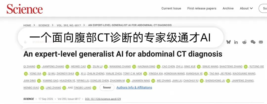
This model can identify 146 diseases across 18 anatomical structures in the abdomen, with higher accuracy than many radiologists.
Moreover, the model, code and framework are all fully open-sourced.
One single model for 146 types of diseases
DAMO RADAR, whose full name is Rapid Abdominal Diagnosis with AI and Radiology, is an AI model for rapid abdominal diagnosis.
This AI "radar" is developed by Alibaba Damo Academy. In the past few years, the R&D team has used AI to identify early cancer lesions that are hardly visible to the naked eye in CT scans, made major breakthroughs in specific diseases such as pancreatic cancer, gastric cancer and colorectal cancer, and published 5 papers in Nature Medicine.
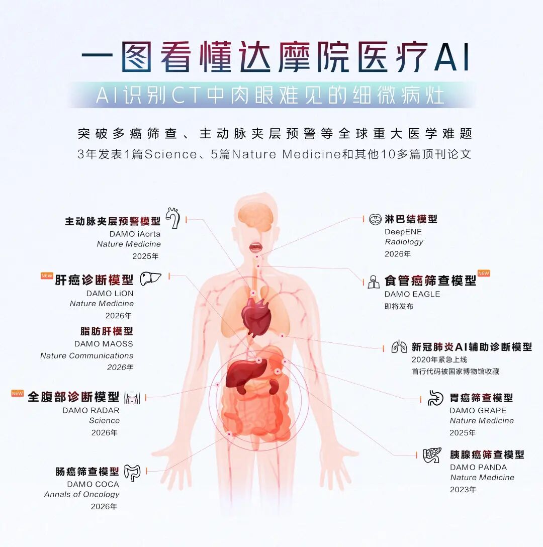
But as the research went on, some members of the Damo Academy team found something wrong. Zhang Jianpeng, senior algorithm expert at Damo Academy, said: "It takes at least two or three years to conquer one disease, and there are tens of thousands of human diseases in total, which means we can never finish all of them in our entire career..."
More tricky is that in the real world, patients do not come to the hospital with only one disease, and the dedicated disease model can only check whether the patient has that specific disease. In fact, one CT image very likely contains multiple organs and multiple lesions at the same time.
Damo Academy joined forces with The First Affiliated Hospital of Zhejiang University and many other top hospitals around the world to tackle abdominal CT imaging, aiming to enable AI to identify all kinds of abdominal diseases at one go.
The abdomen was chosen as the target because it is clinically recognized as the most challenging imaging scenario.
Xiao Wenbo, director of the Department of Radiology of The First Affiliated Hospital of Zhejiang University, introduced that junior doctors are usually most afraid of being assigned to the abdominal imaging group.
The abdomen involves the digestive system, urinary system and reproductive system, with the liver, gallbladder, pancreas, spleen and intestines all crowded together. The intestines coil through the entire abdominal cavity, and almost all organs are soft tissues with similar density, so doctors have to rely entirely on their naked eye's sensitivity to density differences to identify lesions.
Previously, there have been AI auxiliary tools for scenarios such as pulmonary nodules, but the abdomen is far too complex, which has long been a restricted area that no one dares to touch.
What RADAR aims to achieve is covering the entire abdomen with one single model.
Finally, it covers 18 anatomical structures and 146 diseases, including major organs such as the liver, pancreas, gallbladder, kidney, spleen and intestines, basically covering all common clinical lesions.
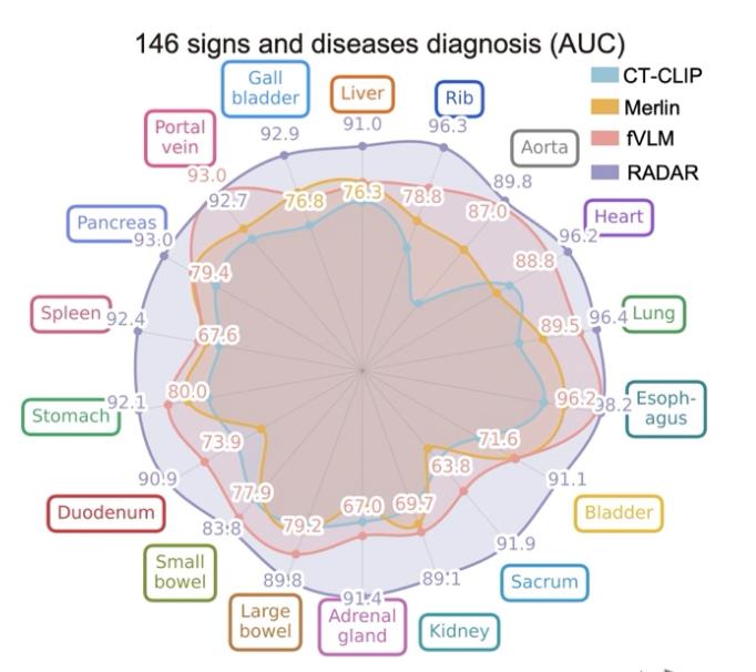
The team conducted very rigorous multi-layer verification on its performance.
In an internal real-world consecutive cohort of nearly 39,000 cases, RADAR's average AUC reached 0.913.
AUC is an indicator that measures the diagnostic model's ability to distinguish patients from healthy people: 1 means perfect discrimination, 0.5 is equivalent to random guessing, and a value above 0.9 is considered excellent.
After that, the team verified the model on more than 24,000 CT scans from 8 external hospitals. These cases cover different regions and different scanning devices, and RADAR's AUC still reached 0.895.
Moreover, RADAR also achieved good generalization performance in emergency scenarios.
The difference between the emergency scenario and the general outpatient scenario is that due to time urgency, there is not enough time for the contrast agent to reach the optimal effect, and patients may not be able to cooperate fully, resulting in suboptimal CT imaging quality.
RADAR's training objectives did not include emergency cases, but when the team tested it with 27,000 emergency data cases, the AUC still reached 0.904.
Furthermore, Damo Academy raised the performance requirements for RADAR in the diagnosis of four types of cancers: liver cancer, pancreatic cancer, gastric cancer and colorectal cancer.
For these four cancers, the team used the "pathological gold standard" as the Ground Truth, that is, taking the pathological biopsy result as the reference to confirm the accuracy of the imaging diagnosis.
In this more demanding test, RADAR's AUC still reached 0.891-0.984.
Damo Academy also conducted a series of human-machine comparison tests.
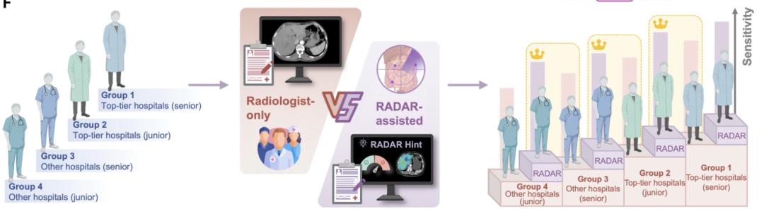
A total of 26 radiologists from 14 hospitals participated in this comparison. RADAR and the doctors made independent diagnoses on the same batch of cases. The results showed that RADAR outperformed 23 of the 26 doctors in accuracy, and was only slightly inferior to the remaining 3 senior doctors.
At the same time, Damo Academy also tested the human-AI collaboration scenario, and found that when doctors read images with the help of AI, the overall sensitivity increased by about 10%, and the average reading time was reduced by more than 30%.
The configuration of "junior doctor + AI" even exceeds the sensitivity of senior doctors reading images alone.
So how on earth did RADAR achieve this?
Traditional medical imaging AI uses supervised learning, which in simple terms requires doctors to label CT images one by one, and then AI learns according to the labels.
This method has extremely high labeling costs, and the model can only identify the few diseases that have been labeled, and cannot recognize other diseases.
RADAR took a different path, called vision-language contrastive learning.
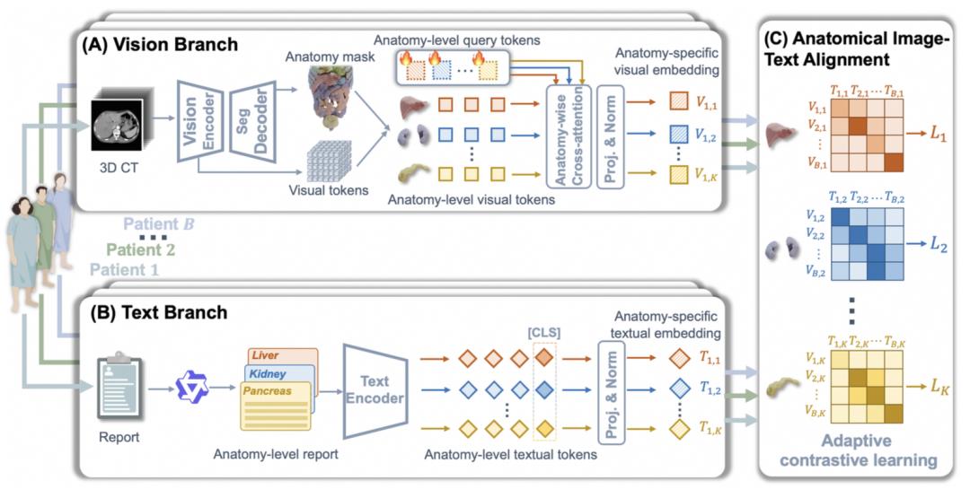
A large number of CT images and corresponding diagnostic reports naturally exist in hospitals, and RADAR learns directly from the correspondence between images and report texts, without requiring doctors to perform additional labeling work for each slice.
This is a bit like a medical student following the attending doctor on ward rounds: when the attending doctor reads the scan, he will say "there is a low-density lesion in the liver, which is considered a hemangioma". After hearing this for many times, the student will naturally know what imaging features correspond to what diagnosis.
However, directly aligning the entire CT scan with the entire report yields very poor results.
The team tried this approach at the very beginning, and found that the model could only learn some rough statistical features, and could not establish the association of "which organ corresponds to which part of the description".
A full set of abdominal CT scans contains hundreds of slices, covering many organs, and the problematic part may only be a tiny lesion on a certain organ. If the model tries to understand the entire image, the key signal will be overwhelmed by a large number of normal tissues.
This leads to RADAR's core breakthrough — "organ-level fine-grained alignment".
It first locates various organs such as the liver, pancreas, gallbladder and kidney from the CT scan, splits the entire set of CT data into individual organ units, and then precisely aligns each organ unit with the corresponding description in the report.
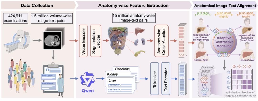
The inspiration for this design comes from the real working pattern of radiologists.
When radiologists read abdominal CT scans, they do not glance at the entire abdomen as a whole. Instead, they check the liver, pancreas, biliary tract, kidney and intestines one after another, processing each organ individually. RADAR allows AI to learn in the exact same way.
Another innovation is called adaptive contrastive modeling.
Standard contrastive learning strictly distinguishes the features of different patients by default, but in medical scenarios, if two people both have healthy livers, their features should not be separated from each other.
Similarly, the imaging features of two patients suffering from the same disease should also be learned together.
RADAR dynamically adjusts the distance between samples based on this medical knowledge.
Moreover, the Scaling Law curve shown in the paper demonstrates that as the data volume increases, the model's performance continues to rise, with no sign of saturation on the curve.
Helping doctors "miss fewer lesions"
The Scaling Law curve is still rising, but the capabilities RADAR has already achieved are changing the working rhythm of frontline doctors.
"When I first saw the verification data of RADAR, I couldn't believe it," said Xiao Wenbo, director of the Radiology Department of The First Affiliated Hospital of Zhejiang University. "AI can detect 146 diseases at one go among so many similar structures in the abdomen, with an AUC of around 0.9, which is completely beyond the expectation of all doctors."
The whole department got excited, many doctors said: Deploy it as soon as possible! Many doctors from other hospitals also heard about this achievement and came to ask for help, hoping that AI can help them solve the tricky abdominal CT problem.
Xiao Wenbo has been working in the imaging field for more than 30 years, leading a department of more than 300 people that processes thousands of scans every day.
Writing a full abdominal CT report takes at least 20 minutes even for senior doctors, as they need to go through hundreds of images in one CT case one by one.
She knows exactly how difficult this work is.
It was not until she found that RADAR maintained a stable high accuracy after repeated rounds of verification that she was truly convinced.
There is a saying in the imaging department: "Same disease with different manifestations, different diseases with the same manifestations". The same disease may present in completely different forms, while different diseases may look exactly the same.
Xiao Wenbo admitted that a doctor may need to read hundreds of thousands of scans to see all kinds of features, and may not be able to access such a large number of cases even in their entire career.
This is precisely the greatest value of RADAR: it fills the blind spot of human doctors and helps them miss fewer lesions.
Especially in life-and-death scenarios such as distinguishing between benign and malignant lesions, doctors are under huge pressure; with AI, it is equivalent to having an imaging assistant at hand at all times, providing a supplementary reference when doctors are hesitating.
However, AI assistance also brings some hidden concerns.
This is not about accuracy, after all, even with AI, there will always be real doctors checking the results at the final step.
The concern is related to the development of future generations of doctors.
Doctor training emphasizes the cultivation of imaging logical thinking, which is polished repeatedly from theory to practice, and this process cannot be skipped.
If junior doctors use AI to write reports, their reports may reach the level of senior doctors. Once they form path dependence, the normal training path will be interrupted.
But there is no need to worry too much.
Xiao Wenbo's idea is to turn RADAR into a "one-on-one teaching tool" at the same time.
She said that the 300+ people in her department are distributed in multiple campuses, and the traditional mode where senior doctors sit next to juniors and guide them through each scan one by one is rarely seen now.
But AI can take on this role: after junior doctors finish writing the report as usual, they can use AI to conduct a self-check, and immediately find out which lesion they missed and which judgment is biased.
It is just like having a senior doctor standing by and saying, "Hey, there is another lesion here, why didn't you notice it?"
In this way, every case becomes a process of cross verification and learning.
Looking further ahead, RADAR's organ-level alignment framework is also compatible with other imaging modalities. As long as relevant data is available, MRI, PET and ultrasound can all become the targets for migration.
Moreover, Damo Academy has not kept these achievements private: the model, code and technical framework are all fully open-sourced, and any team can reproduce, improve and migrate it to their own scenarios.
At this point, this story is far more than just a story about a medical AI.
The idea behind RADAR is to use one single general-purpose model to solve problems across an entire field.
Medicine is the scenario with the most extreme accuracy requirements and the lowest error tolerance among all industries. One misjudgment may change a patient's treatment plan or even their survival time.
If this paradigm can work in this scenario, it can most likely be applied to other high-precision scenarios as well.
Zhang Ling, senior algorithm expert at Damo Academy, said: "Science very rarely publishes papers on medical imaging AI, because it has long been regarded as an engineering technical problem. RADAR proves for the first time that general-purpose medical imaging AI is a technically feasible route, which changed the mind of Science's reviewers, who began to treat it as a 'scientific problem'."
The fact that RADAR passed this strict threshold proves that this method has withstood the