The reshuffle of the ophthalmic equipment industry: Can one single device handle all examinations? The ambition of domestic integrated solutions
Rows of ophthalmic examination devices are lined up, with patients leaning forward in front of each one sequentially, moving to the next unit after completing a single test. Doctors constantly shuttle between devices, unable to assign one dedicated device to each patient. In the ophthalmology examination areas of many hospitals, this assembly-line-style workflow repeats busily every day.
Integrated devices that consolidate multiple functions into a single unit can streamline processes or improve diagnosis and treatment accuracy, becoming a key direction for optimizing this ophthalmology "assembly line".
In recent years, the development of domestically produced high-end ophthalmic devices has accelerated, with products such as OCT, optical biometers, and ultra-wide-angle fundus cameras seeing rising market share. However, the core positions in the ophthalmology departments of large hospitals are still occupied by imported devices, which can handle multiple tasks on their own, significantly reducing the burden on doctors and departments.
Nowadays, with the successive launch of domestically produced integrated products such as multimodal imaging devices, intraoperative navigation microscopes, and combined phacoemulsification and vitrectomy systems, the localization of high-end ophthalmic devices has entered a new, more competitive stage.
01 Further Evolution of Multimodal Diagnostic Devices
The human eye has a multi-layered three-dimensional structure with complex anatomy, so ophthalmic diagnosis and preoperative evaluation rely on data collected by various types of devices. Different imaging and measurement technologies have distinct principles and advantages, and the complementary strengths of multiple technologies can present ocular tissue morphology, visual function, and lesion conditions from different dimensions.
Taking fundus imaging as an example, modalities such as color fundus photography, infrared scanning imaging (IR), fluorescein angiography (FFA), indocyanine green angiography (ICGA), and optical coherence tomography (OCT) have become commonly used techniques.
However, in the past, the devices corresponding to various technologies were independent of each other. Patients needed to move between multiple instruments to complete a comprehensive examination, and departments also had to assign multiple full-time technicians, resulting in cumbersome procedures and high labor costs. Separate shootings would cause spatiotemporal deviations, making it difficult for different images to accurately match the same lesion, thus affecting precise diagnosis.
With the development of laser diagnostic technology, laser scanning ophthalmoscopes that integrate multiple imaging modes based on multi-wavelength laser imaging capabilities have become typical integrated diagnostic devices. Among them, imported brands such as Optos, Heidelberg, and Nidek have extensive clinical recognition based on years of accumulated fundus imaging technology. In recent years, domestic enterprises including ClearVue Medical, Yaoshi Medical, Bigway Medical, and Topmed Medical have also successively launched similar devices: laser scanning ophthalmoscopes that integrate imaging modalities such as ultra-wide-angle color photography, autofluorescence, and angiography.
More importantly, the current trend of fundus imaging integration has further escalated: the deep integration of laser scanning ophthalmoscopes and OCT has become a new direction. In this setup, planar images such as color fundus photography present the full view of the fundus, while OCT analyzes the three-dimensional tomographic structure of deep ocular layers, making the two a natural perfect pair.
While several major international brands are advancing in this integration direction, domestic devices have also achieved technological integration breakthroughs, especially successfully realizing one-click simultaneous multimodal imaging.
For example, the ClearVue Medical SKY Full-Eye Imaging Platform integrates two cutting-edge fundus imaging technologies: confocal laser scanning (CSLO) and swept-source optical coherence tomography (SS-OCT), supporting core functions such as ultra-wide-angle laser color photography, laser angiography, and ultra-wide and ultra-deep swept-source OCT, with X types of corresponding images that can be collected simultaneously.
"When using traditional multimodal devices in connected mode, there are still situations where lenses need to be replaced and functions switched. Patients have to undergo separate examinations for each item, and multimodal images of the same lesion still have angular deviations, which may affect accurate diagnosis," introduced Xu Haijun, Marketing and Sales Director of ClearVue Medical. After overcoming challenges such as optical path interference from multi-channel simultaneous laser scanning and AI algorithms for planar and tomographic image registration, the SKY Full-Eye Imaging Platform achieves one-click multimodal imaging, allowing doctors to accurately observe the lesion location of patients at the exact same time and spatial position, which is particularly critical for identifying tiny lesions.
For instance, in the diagnosis of complex macular diseases, this imaging mode can automatically locate the macula, quantify the area of fluid accumulation or atrophy in layers, and distinguish between dry and wet macular degeneration; in glaucoma diagnosis, it can simultaneously compare the optic disc morphology and optic nerve thickness, using unified standards for planar and tomographic image comparison to reduce manual positioning errors.
The deep integration of OCT and optical biometry is another direction for the integration of ophthalmic diagnostic devices. Intalight Saiwei's OCT, as well as optical biometers from enterprises such as Topmed Medical, Moptim Medical, Bigway Medical, and Weiren Medical, integrate OCT and biometry into one unit: the former is responsible for visualizing structures, while the latter performs precise measurements, providing one-stop accurate examinations for myopia prevention and control or surgical procedures.
Overall, technical routes such as multimodal integration, the unification of planar imaging and tomographic devices, and the fusion of imaging and measurement have become important trends in the iteration of ophthalmic diagnostic devices. Leveraging differentiated advantages such as one-click simultaneous imaging, automated operation, and adaptability to primary medical institutions, domestic devices continue to narrow the technological gap with overseas products, providing complete solutions for hospitals at all levels to build an efficient, accurate, and low-cost fundus diagnosis and treatment system.
02 Breakthroughs in High-Technology Surgical Devices One After Another
For a long time, domestically produced ophthalmic therapeutic devices have been a weak point. In the past, domestic devices were mostly concentrated in categories such as meibomian gland physiotherapy, intense pulsed light, laser photocoagulation, and mid-to-low-end ophthalmic surgical microscopes, with relatively single functions, while multi-functional complex surgical devices were monopolized by overseas brands for a long time. Now, with the approval of domestically produced combined phacoemulsification and vitrectomy systems and intraoperative navigation microscopes, the gaps in integrated surgical devices are being filled.
In reality, many cataract patients have combined fundus lesions such as retinal or macular disorders. If separate phacoemulsifiers and vitrectors are used for staged surgeries, two anesthesia procedures are required, which not only increases the risk of wound infection but also prolongs the treatment cycle, increasing the financial, physical, and mental burden on patients. As an integrated surgical platform for the anterior and posterior segments, the combined phacoemulsification and vitrectomy system can precisely solve these pain points. Before 2025, the domestic market in this track was entirely occupied by imported brands such as Alcon and Bausch + Lomb.
The vitrectomy module is the difficulty in developing the integrated system, with technologies covering interdisciplinary fields such as gas-liquid management, dual-pump fluid regulation, and real-time stable negative pressure feedback. The overall system has complex mechanical, electrical, and embedded system architectures, with long debugging cycles and huge R&D investment. While mastering these technologies, imported brands continue to make core upgrades around higher cutting efficiency and safer intraocular pressure control.
Filling the domestic gap not only requires matching the hardware performance of imported devices but also building differentiated advantages based on domestic clinical needs.
From 2025 to the present, two domestically produced combined phacoemulsification and vitrectomy systems have been successively approved for launch, with core functions and performance comparable to imported devices.
The VitroRx Medical Genesis series products have vitrectomy cutting speeds of 15k and 20k CPM respectively, capable of performing all types of vitrectomy surgeries from 20G to 27G; the phacoemulsification system uses a 28.5kHz frequency and a 1.8mm incision to reduce postoperative iatrogenic astigmatism and incision thermal damage; in addition, the products also feature light source wavelength switching and ultrasonic vitrectomy functions.
Based on full-stack self-developed core technology platforms for fluidics, optics, and energy, the SIERRA VISION SYSTEM® integrated anterior and posterior segment surgical platform from Siora Medical integrates core functions such as phacoemulsification, vitrectomy, laser, electrocoagulation, and illumination, capable of completing the entire workflow from cataract phacoemulsification to vitrectomy, laser photocoagulation, and intraocular filling without the need for additional external devices; it is equipped with China's first domestically developed 20K CPM ultra-high-speed double-blade dual-airpath inclined vitreous cutter, with high cutting efficiency and laser transmission efficiency of over 90%.
The integrated system is not a simple superposition of devices, but a closed-loop system including the main unit, intelligent software, and supporting consumables. In addition to core hardware parameters, the overall system performance also relies on dedicated consumables and regular maintenance support. Domestic devices build differentiated advantages in aspects such as site adaptation, consumable usage, and local supply chains, forming full-lifecycle solutions to reduce costs and increase efficiency for medical institutions.
For example, the VitroRx Medical Genesis series adopts a modular design, facilitating rapid upgrades and iterations in the later stage; it develops exclusive disposable consumables, which will further reduce the total cost of the system plus consumables as clinical large-scale adoption progresses in the future.
The Siora Medical SIERRA VISION SYSTEM® is equipped with an upgraded dual-LED light source, with a single light source having a service life of up to 30,000 hours. Compared with the xenon lamps commonly used in imported devices that only last for hundreds of hours, there is no need to frequently replace light source consumables throughout the entire usage cycle; the overall system footprint is reduced by 30%, and the lightweight design adapts to operating rooms with limited space.
When talking about other advantages of localization, Liu Kunchi, Marketing Director of Siora Medical, also stated that on the basis of fully independent and controllable R&D, production, and supply chains, domestic devices can be produced and delivered locally for both the main unit and supporting consumables, and can also respond faster to medical institutions' maintenance needs, continuously ensuring stable device operation.
While domestic breakthroughs have been achieved in the integrated surgical platform, the gap in domestically produced intraoperative navigation microscopes has also been filled.
This device integrates a surgical microscope with intraoperative OCT, displaying real-time fundus tomographic images during surgery, making it easier for doctors to identify lesions and reduce operational risks. Its R&D involves cutting-edge technologies such as high-precision optics and image synchronous fusion. Before 2024, there were only two overseas brands, Zeiss and Leica, in the domestic market.
Dual optical path fusion is the core barrier for intraoperative navigation microscopes. The navigation microscope launched by Topmed Medical in 2025 adopts a coaxial integrated design of the OCT and the main optical path of the microscope, completing the deep integration of the two technologies. The optical path structure and the number of components have increased significantly, solving the pain points of lagging images and narrow fields of view from previous externally mounted OCT systems, better meeting the clinical needs for real-time perspective observation.
Overall, domestic breakthroughs in high-end ophthalmic surgical devices have moved from the stage of catching up with single-function devices or consumables to a stage of integrated systematic innovation.
03 Where Will Device Integration Go?
Various signs indicate that domestically produced integrated ophthalmic devices have officially stepped onto the industry stage:
Since its launch over a year ago, the ClearVue Medical SKY Full-Eye Imaging Platform has not only been deployed in medical institutions at all levels across China but also sold to overseas markets. The Siora Medical SIERRA VISION SYSTEM® was approved in April 2026, and just three months later, several units had already won bids in public hospital procurement projects. The price of Topmed Medical's intraoperative navigation microscope is comparable to similar imported high-end models.
In addition to products that have entered the commercialization stage, domestic enterprises have also laid out a rich pipeline of under-development integrated ophthalmic devices.
"There is a long-term demand for functional integration of devices in ophthalmic operating rooms. Clinical applications such as combined anterior-posterior segment surgery and combined cataract-glaucoma surgery are becoming more and more widespread. Integrated devices, along with their intelligence and visualization capabilities, are the inevitable direction to meet these surgical needs. Primary medical institutions generally face budget and space constraints, so a single device that covers multiple types of diagnosis and treatment scenarios can effectively reduce daily operational pressure," analyzed Liu Kunchi.
In the short term, intraoperative navigation microscopes and combined phacoemulsification and vitrectomy systems will remain the main types of product R&D.
Surgical microscopes are the basic configuration for ophthalmic surgeries. In the past, products were mainly based on traditional optical and mechanical technologies. With the continuous integration of multimodal imaging and intelligence in this field, the demand for hospital device replacement continues to be released. Currently, enterprises including Siora Medical, VitroRx Medical, Moptim Medical, and New Optics have already started R&D layouts.
As the population aging in China continues to deepen, the demand for combined surgeries for cataract with fundus lesions is increasing year by year, giving the combined phacoemulsification and vitrectomy system huge market potential. It can not only replace the current mainstream separate phacoemulsifiers and vitrectors in the existing stock market but also tap into the incremental market. Corresponding products from enterprises such as Topmed Medical, New Eye Medical, and Jiasinuode Medical are all in the R&D stage.
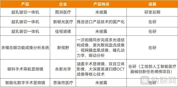
Some under-development integrated ophthalmic devices, Source: Public information from enterprises
In addition, ophthalmic surgical robots and femtosecond lasers are also typical integrated devices, and the first domestically produced unit is about to be released.
Looking at the longer term, integrated ophthalmic devices will continue to iterate and upgrade around actual clinical needs, deriving more diverse functional integration solutions and product forms.
Xu Haijun believes that these integrated examination devices will see explosive market demand in the future: first, integrated devices for large-scale school screenings that combine multiple functions such as optometry, intraocular pressure measurement, fundus photography, and strabismus and amblyopia screening; second, integrated preoperative evaluation systems for refractive surgery that integrate full sets of preoperative detection modules for the cornea and fundus. These two types of devices can effectively improve the examination efficiency and diagnosis accuracy of medical institutions.
In terms of product form, compact and mobile integrated devices will become increasingly popular in the sinking market. For scenarios such as community health service centers, township health centers, and mobile medical examination vehicles, lightweight multimodal integrated systems will become the R&D focus; a single device can simultaneously complete optometry, intraocular pressure detection, wide-angle fundus imaging, and OCT scanning, without requiring large supporting venues, extending efficient eye health screening services to towns and communities.
At the same time, relying on multimodal data interconnection technology, the industry will realize integrated image management throughout the entire process of screening, surgery, and reexamination. At this stage, separate devices generally face the problem of image data silos, where imaging parameters and lesion positioning coordinates of different devices cannot be uniformly interconnected; in the future, integrated devices and platforms will connect data across all links: preoperative image examination, intraoperative OCT navigation, and postoperative reexamination. Doctors can access the complete cycle of a patient's ocular image data to continuously track disease changes, which is especially suitable for long-term follow-up management of chronic eye diseases such as diabetic retinopathy and glaucoma.
It is not difficult to see that the key for domestically produced high-end ophthalmic devices to increase market share is not just "copying homework" to replace imported products, but also to align with the needs of China's medical system, deeply understand clinical scenarios, and create products with excellent performance and stronger adaptability. Leveraging the good start of existing products, domestically produced ophthalmic devices will continue to integrate cutting-edge technologies in the future, competing head-to-head with international brands in the global market.
This article is from the WeChat Official Account "Arterial Network" (ID: vcbeat), Author: Zhang Xiaoxu, published with authorization from 36Kr.