Can AI really determine a person's life and death through face reading?
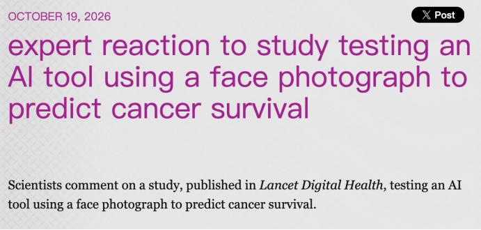
On October 20, the international medical journal *Lancet Digital Health* published a study: AI can identify differences in mortality risk among cancer patients using only an ordinary facial photo.
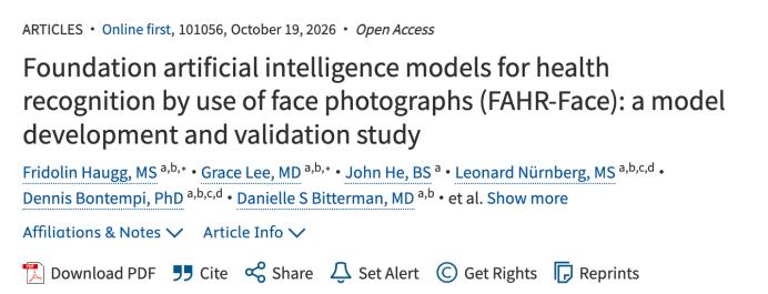
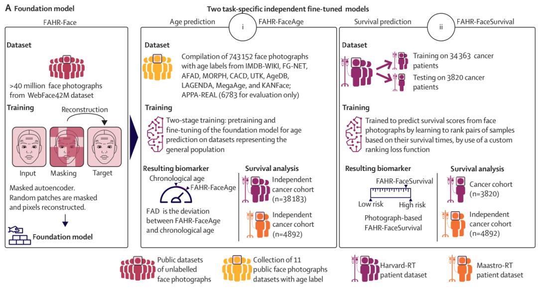
Mass General Brigham developed an AI model named FAHR-Face, which is trained to find clues related to patients' survival conditions from facial photos.
The researchers recruited 3820 cancer patients who had not participated in model training for testing, among them, 122 were rated as extremely high risk by AI, and 64 of these 122 people died within 90 days after the photo was taken.
The researchers then conducted external validation using data from nearly 4900 Dutch cancer patients, and similar results were observed.
Throughout the whole prediction process, the AI does not need to check medical records, analyze CT images, or refer to blood test results. It only needs to look at a face.
01
AI can predict mortality risk with just one face photo
The most critical point of this study is that AI can truly distinguish cancer patients with different mortality risks.
To achieve this, the research team first used more than 40 million ordinary facial photos to train a visual foundation model called FAHR-Face.
The training method is a bit like a jigsaw puzzle: the researchers randomly covered about 75% of the area in the photo, and then let the AI try to restore the complete photo based on the remaining part. After repeated training, the AI gradually learned to recognize the structure of human faces and the connections between different facial features.
With this foundation, the researchers began to teach it to predict mortality risk. They collected 34363 facial photos of cancer patients, as well as corresponding survival records, including whether the patient had passed away, the time of death, or how long the patient had survived by the last follow-up.
Next, the researchers adopted a ranking learning method to let the AI compare the survival time of different patients. For example, after two patients took photos, one died within three months and the other survived for a longer time. The model needs to try to assign a higher mortality risk score to the former.
Through a large number of such comparisons, the AI gradually learned to judge the relative mortality risk of different patients from facial features.
Finally, the researchers converted the scores output by the model to a range between 0 and 1. The higher the score, the higher the mortality risk of the patient as judged by the AI.
To verify the accuracy of the model's scores, the researchers additionally recruited 3820 cancer patients who did not participate in the training.
As a result, in addition to the 122 extremely high-risk patients mentioned at the beginning, the AI also assigned risk scores between 0 and 0.1 to another 975 patients. Among these 975 people, only 20 died within 90 days after taking the photo, accounting for only 2.1%.
In contrast, the 90-day mortality rate of extremely high-risk patients reached 52.5%, with a difference of more than 25 times between the two groups. The high and low risk scores given by the model do correspond to completely different mortality rates.
Moreover, this difference does not only exist in the two extreme intervals with the highest and lowest risks. The researchers divided the patients into four groups from low to high according to the risk scores given by AI, and found that as the scores increased, the survival rate of patients also decreased significantly.
Even after incorporating factors such as age, gender, cancer type, and treatment status into the statistical analysis, the mortality risk of the highest-risk group was still 3.23 times that of the lowest group.
However, there is still a problem at this point: this model performs well in US hospitals, can it still get similar results when applied to another hospital and a new group of patients?
The researchers recruited another 4892 patients from the Maastro Radiation Therapy Center in the Netherlands and tested them using the same model.
As a result, they again observed a similar pattern: the higher the score given by AI, the higher the overall mortality risk of patients. Moreover, this difference still exists after taking factors such as age, gender, and cancer site into account.
From the test data of a hospital in the United States to the independent patient cohort of another hospital in the Netherlands, this model can identify significant differences in mortality risk among cancer patients.
So, what exactly did the AI see on the faces of these patients?
02
What exactly did the AI see from human faces?
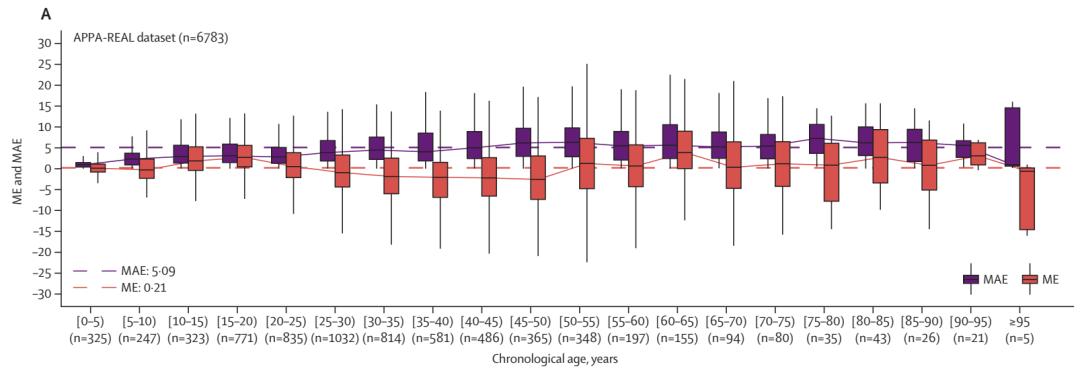
In fact, FAHR-Face is not limited to the mortality risk prediction function mentioned earlier. The research team also developed another model called FAHR-FaceAge, which is specially used to estimate a person's facial age.
The idea of this model is relatively easy to understand: even for people of the same actual age of 60, some look like they are only in their early 50s, while others look much older. The researchers believe that how much older a person looks than their actual age may also reflect their health status.
They used hundreds of thousands of facial photos labeled with age to train the model, and then calculated the gap between the facial age estimated by AI and the actual age. The results showed that among cancer patients, those who looked older than their actual age often faced higher mortality risk.
For example, after taking factors such as age, gender, and cancer type into consideration, patients whose facial age estimated by AI is at least 5 years older than their actual age have a mortality risk about 21% higher than other patients.
But the research team did not stop there.
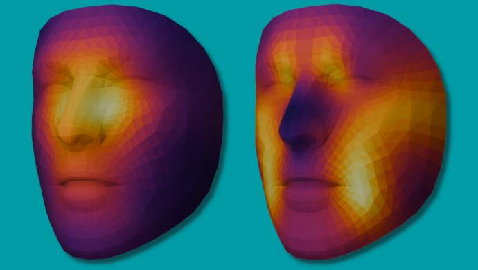
They wanted to know whether there was other information related to mortality risk hidden on human faces, apart from how old a person looks. Therefore, they analyzed the age model and the mortality risk model respectively, and drew two sets of attention heatmaps for the AIs.
The results showed that the areas they focused on were significantly different.
When predicting age, the AI mainly focuses on the nasolabial folds, the forehead and the area near the temples; when predicting mortality risk, it pays more attention to the area under the eyes and around the bridge of the nose.
The researchers also used statistical methods to compare the two models. Among 4892 patients in the Netherlands, the correlation coefficient between the gap between facial age and actual age and the mortality risk score given by AI was only 0.16, which means the correlation between the two is very weak.
Moreover, when the researchers combined the two indicators to predict the survival risk of patients, both of them retained independent predictive effects, and the performance of the combined statistical model was also improved.
In other words, from the same facial photo, AI can extract two different but complementary sets of risk prediction information.
But here comes the question: what exactly is this information?
The research team proposed in the paper that the features under the eyes and around the nose may be related to the facial changes of severely ill patients that doctors have observed for a long time. For example, when the patient's body gradually weakens, the state of facial fat, muscles and skin may all change, and these changes may leave traces on the photo.
However, the researchers have not yet proved what specific physiological changes the AI has identified.
Miroslaw Bober, a professor of machine learning at the University of Surrey in the UK, also pointed out this problem in an expert comment released by the UK Science Media Centre. He believes that this study does prove that there is a statistical correlation between patients' facial features and survival risk, but being able to predict the outcome does not mean that the cause has been figured out.
Speaking of which, this reminds people of the "observation, auscultation and olfaction, inquiry, and palpation" that traditional Chinese medicine emphasizes. The "observation" part refers to looking for clues related to health status by observing a person's facial color, expression and body shape. In modern medicine, doctors also pay attention to whether patients have physical appearance changes such as emaciation and haggardness.
Interestingly, the paper of this study even specifically cites medical literature on "Hippocratic face" — "Hippocratic face" refers to the special facial features that some critically ill patients, especially those on the verge of death, may present: sunken eye sockets, sunken temples, sharpened nose, pale or dull complexion. More than 2,000 years ago, Hippocrates recorded these features and used them as clues to judge whether a patient's condition was critical.
Nowadays, AI is trying to turn the observations that doctors used to make based on experience into quantifiable risk indicators.
But there is still no answer as to whether it has discovered new clues that are difficult for human eyes to detect, or has converted the physical changes that doctors could originally observe into digital values.
03
How much medical information is hidden in a face?
Interestingly, some of the photos used by the researchers are just ordinary photos usually taken by hospitals to verify patients' identities.
This is not their first time to conduct research on facial photos.
In May 2025, the team published their initial FaceAge study in *Lancet Digital Health*. At that time, they also tried to let AI assist doctors in predicting the survival time of patients with advanced cancer.
The researchers recruited 100 patients receiving palliative care, and asked doctors and researchers to judge whether they would still be alive half a year later. After adding the information provided by FaceAge, the prediction performance was improved: AUC, an indicator that measures predictive power, increased from 0.74 to 0.80.
In this new FAHR-Face study, researchers have begun to try to directly predict mortality risk from photos.
The most attractive part of this research route is that it is extremely convenient.
CT and MRI require special equipment, and pathological examinations require tissue samples to be collected, while facial photos only require an ordinary camera. For doctors, this means that there may be a low-cost, easy-to-operate auxiliary assessment method in the future that will not add physical burden to patients.
Raymond Mak, the lead researcher and a radiation oncologist, mentioned that when doctors decide whether to arrange surgery, radiotherapy or other high-intensity treatments for patients, they need to assess the patient's physical condition and whether they can tolerate the treatment. He hopes that AI can make such judgments more objective and unified in the future.
However, there are still many problems to be solved before this goal can be truly achieved.
First of all, although ordinary photos are easy to obtain, they are also easily affected by shooting conditions. Professor Miroslaw Bober from the University of Surrey in the UK pointed out that just asking a person to smile, or changing the light when taking a photo, may cause the facial age estimated by AI to change by an average of about two years, and sometimes the difference will be even greater.
Although this does not mean that the mortality risk model will be affected in the same way, if we want facial photos to become a reliable medical indicator, we must first figure out how much interference these shooting conditions will cause.
In addition, the photos of cancer patients used in current research are still mainly of white people. For other populations, more data verification is needed to confirm whether the model can maintain the same predictive performance.
More importantly, predicting mortality risk is still not the same as truly helping doctors treat patients. The 2025 FaceAge study has shown the potential to assist doctors in making judgments, but this new model has not yet fully proved how much additional help it can provide on the basis of doctors' existing clinical assessments.
It can divide patients into different groups according to mortality risk, but it has not yet proved whether these predictions can help doctors choose a more appropriate treatment plan. Therefore, FAHR-Face cannot be directly used in clinical practice for the time being.
By the way, Mass General Brigham is not the only institution trying to find disease clues from ordinary facial photos.
As early as 2020, researchers from institutions including Fuwai Hospital of the Chinese Academy of Medical Sciences and Tsinghua University published a study in *European Heart Journal*, trying to let AI identify coronary heart disease through facial photos.
In tests on 1013 patients, the model achieved an AUC of 0.73, showing certain disease recognition capabilities.

In February this year, the research team from institutions including the University of Copenhagen in Denmark focused on the emergency room. They collected mobile phone photos of 27660 emergency patients and let AI predict the mortality risk of these patients.
The results showed that with only facial photos, AI can identify some high-risk patients who are easily overlooked by traditional emergency triage systems. Some people who were originally considered to have mild conditions were rated with high mortality risk by AI, and they were indeed more likely to die later.
However, this study is currently only a preprint and has not undergone formal peer review.
From cancer to coronary heart disease, and then to emergency patients, a number of studies have proved that an ordinary facial photo does contain information related to the patient's health, and this information may be richer than we previously imagined.
As for what role this information can play, perhaps researchers need to continue to find the answer.
This article is from the WeChat Official Account "Alpha AI", author: Yuan Xinyue, editor: Wang Jing, published with authorization from 36Kr.