Using AI to customize "brain maps" for patients, Xijia Medical has completed tens of millions of yuan in Pre-A round financing | 36Kr Exclusive
By | Lingguo Liu
Edited by | Ruojing Hai
36Kr learned that Xijia Medical, an AI brain science company, recently completed a Pre-Series A financing of tens of millions of yuan, led by Sinocapital, with participation from Xi Venture Capital, Qinzhi Capital, Huichuang Medical and Xingfu Capital. The proceeds from this round of financing will be mainly used for clinical research, market expansion and product iteration.
Founded in 2019, Xijia Medical focuses on analyzing MRI (Magnetic Resonance Imaging) data through AI and cloud computing technologies combined with connectomics, so as to generate accurate and individualized brain network maps for patients. At present, this technology is mainly used for neurosurgical surgical planning, neuromodulation, and pre-implantation positioning of electrodes for brain-computer interfaces.
In May 2025, Xijia Medical disclosed an angel round financing of tens of millions of yuan, invested by Xianfeng Qiyun Investment, which was mainly used for the market promotion and further R&D of AI connectomics products. More than a year has passed, and this investment has yielded initial results.
It is understood that based on NuraTome, its AI brain atlas software, Xijia Medical has further developed TMS (Transcranial Magnetic Stimulation) navigation equipment. Based on its precise target scheme for TMS treatment of depression, the scale reduction rate of treated patients has improved significantly compared with the baseline, showing a good clinical remission rate.
At the same time, the company's business lines have expanded to brain-computer interface scenarios, having cumulatively supported more than 20 cases of clinical electrode implantation, providing upstream navigation infrastructure for brain-computer interface enterprises.
From "Average Brain" to "Individual Coordinates"
In 2016, researchers including Matthew F. Glasser divided the cerebral cortex into 360 brain regions based on the MRI data of the Human Connectome Project (HCP), and released a brain atlas covering information such as cerebral cortex structure, brain function, and regional connections. Since then, as one of the general brain atlases, the HCP atlas has been widely used in brain research and encephalopathy diagnosis and treatment.
However, the HCP atlas mainly presents an "average" brain, and "human head sizes, sulci and gyri are all different. In some cases, tumors and edema will squeeze surrounding tissues and displace brain regions." mentioned Quan Yuan, CFO of Xijia Medical. Therefore, the HCP atlas cannot be directly applied to different patients.
Interventions that access the brain, such as neurosurgery, magnetic stimulation, and electrode implantation, require precise positioning of their structures and functions. How to accurately register the general atlas with different brains? Xijia Medical has provided a new solution to this problem through machine learning on massive MRI data.
Specifically, its core software product NuraTome contains an atlas framework of 379 brain regions, of which 360 cortical brain regions are from the HCP atlas, and 19 additional subcortical regions are supplemented.
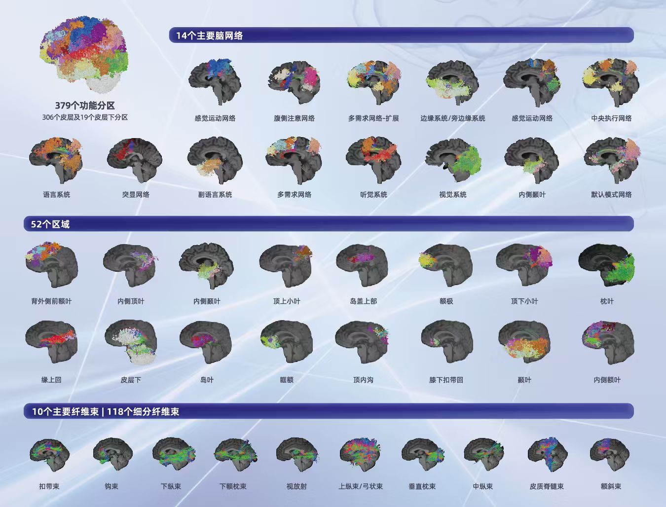
Xijia Brain Atlas - Core Brain Structures
According to the introduction, as early as 2017 before the emergence of large models, the Xijia team began to build AI capabilities for brain imaging, using MRI data from healthy people and patients to train dedicated algorithms. The AI model first learns typical connection patterns of nerve fiber tracts in different brain regions from data of healthy people, and on this basis, further learns to identify the "unique" positions corresponding to each "standard" brain region in different patients' brains. This method of individualized brain network positioning based on structural connections is also called SCA (Structural Connectivity Atlas). On this basis, NuraTome combines functional MRI (fMRI) to analyze functional connections between brain regions.
In the end, what doctors see through the Xijia NuraTome software is an individualized brain network "map" that contains both structural connection spectra and functional connection atlases. Yuan Quan said that Xijia's ecological niche can be analogous to the position of map enterprises in the Internet industry. By this analogy, the brain structure atlas can be understood as delineating roads, buildings, divisions, etc. on the map, while the functional atlas adds dynamic information such as traffic density, traffic lights, and congestion points.
With the coordination of the two, doctors can more accurately determine the location of the lesion, track the direction of fiber tracts, and identify functional sites, so as to determine the intervention method and path.
Yuan Quan said that at present, Xijia has accumulated tens of thousands of domestic and foreign clinical data cases for AI training, in addition to years of scientific research data and public data sets, and the scale of data accumulation is at the forefront of the industry.
Stock data is one aspect. To continuously improve model capabilities, incremental data is also critical, which can make the "speed" of the data flywheel faster. According to the introduction, NuraTome uses resting-state functional MRI, and patients only need to lie still for about 8 minutes to complete the scan. In comparison, traditional task-state scanning may take two to three hours, so Xijia's data accumulation speed on MRI is also relatively faster. "The full 23-minute MRI scan plus 90 minutes of data analysis can complete the data processing."
"Both Software and Hardware Capabilities"
The field of neuromodulation for TMS (Transcranial Magnetic Stimulation) is the business line that has made the most rapid progress for Xijia in the past year.
TMS is a therapy that stimulates or inhibits neuronal activity in specific brain regions through pulsed magnetic fields, mainly including three steps: positioning the target, coil navigation, and magnetic stimulation output. In the past, doctors usually combined images to estimate the target position according to anatomical landmarks, then held the coil by hand to align the patient's head for magnetic stimulation treatment.
"In traditional therapies, taking depression as an example, the structural center point of the dorsolateral prefrontal cortex (DLPFC) is mainly selected as the stimulation target, but this brain region is about the size of a tennis ball, and the stimulation focus of the TMS coil is limited, so in actual treatment, it is difficult to accurately stimulate the effective target; NuraTome can further reduce the positioning range to the millimeter level, telling doctors the specific position to stimulate."
In addition, under traditional therapies, due to the lack of individualized understanding of different patients' brain networks, patients with the same indication are often stimulated at the same target during treatment. But in fact, behind the same indication, the abnormal points of the brain network may be different. For example, for depression, some patients mainly show low mood, some have anhedonia, or are troubled by anxiety, insomnia, etc. Although the scale scores are close, it does not mean that abnormal connections occur in the same brain region. As a result, stimulating the same position will naturally reduce the therapeutic effect. According to the introduction, under the "blind stimulation" mode of positioning based on traditional anatomical landmarks, the effective rate of TMS treatment is sometimes only 20% to 30%, and the clinical benefit for patients is not stable.
To achieve personalized targets for different people, we must first identify the abnormal points in each patient's brain network. In terms of specific mechanism, NuraTome will compare the patient's functional connections with reference data from healthy people, find out the regions where connections are over-active or inhibited, and then combine indications, symptom manifestations, clinical data, etc., to screen out the connections related to the patient's symptoms and form candidate therapeutic targets.
According to the introduction, Xijia has now formed target calculation strategies for about ten types of indications including depression, sleep disorders, anxiety disorders, Alzheimer's disease, autism, ADHD, and post-stroke rehabilitation. The company said that after adopting its precise target scheme, the effectiveness of TMS treatment has been significantly improved compared with traditional methods.
After accurately finding the target, the TMS therapy is only half completed, and the other half is "coil navigation". During the treatment, doctors need to ensure that the coil is always aligned with the target. In the past, this part was mainly completed by third-party navigation hardware equipment, and Xijia was only responsible for software positioning.
According to the introduction, Xijia has now self-developed TMS navigation equipment, integrating positioning and navigation functions into one set of equipment. After NuraTome calculates the target, the binocular camera of the navigator will track the position of the patient's head and the coil, assisting doctors to adjust the coil and reduce deviation during treatment. Yuan Quan uses shooting as an analogy: "TMS is the gun, the navigation equipment is the sight, and NuraTome is responsible for identifying high-value targets."
Find the Landing Point, Draw the Boundary
In addition to neuromodulation, as brain-computer interfaces have accelerated into clinical practice in the past two years, due to their same demand for precise brain positioning, they have naturally become a new scenario for NuraTome to expand.
Whether the invasive brain-computer interface implants electrodes into the brain tissue or attaches them to the dura mater, or the non-invasive brain-computer interface collects signals such as EEG from outside the scalp, it is necessary to capture neural signals to decode human intentions and convert them into equipment instructions.
Whether the signals can be collected clearly enough depends on, on the one hand, the number of electrodes and channels, and on the other hand, the specific position where the electrodes are placed. Specifically, the closer to the signal hotspot, the greater the chance of capturing effective signals.
Taking the invasive brain-computer interface for motor function reconstruction as an example, traditional implantation usually judges the general range of the motor function area according to anatomical landmarks such as the "hand knob area" of the anterior central gyrus of the brain. However, the hand knob area has morphological differences such as Ω shape and ε shape, and the real hand movement hotspots may not completely overlap with it. In this case, NuraTome, with its ability to identify functional hotspots, key nerve fiber tracts and safety boundaries, can refine the electrode target area for doctors and plan the specific implantation path.
According to the introduction, the company has currently cooperated with many leading brain-computer interface enterprises, having cumulatively supported more than 20 cases of clinical implantation, mainly adopting the per-case billing model.
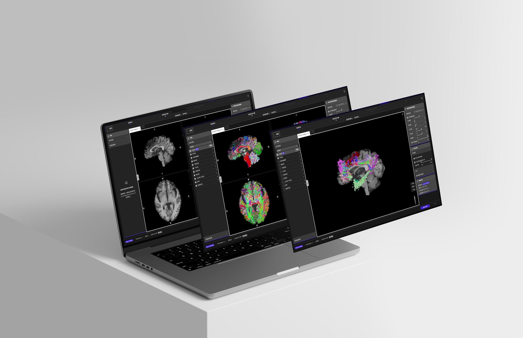
Xijia1x Magnetic Resonance Image Processing Software
The third main application scenario of NuraTome, neurosurgery, is where its positioning capability was first tested.
"Based on our individualized brain atlas imaging, doctors can clearly see the brain network, nerve fiber tracts, and gray matter areas around the tumor, so as to better judge what impact cutting here may have on the patient's brain function." Yuan Quan said.
Cutting one more millimeter too much may hurt normal brain tissue; cutting one less millimeter too little may lead to incomplete tumor resection and subsequent recurrence. The lack of tools to accurately locate the tumor resection boundary is a long-term difficulty faced by neurosurgery. Yuan Quan took a thalamic glioma surgery as an example to explain that this type of tumor is located deep in the brain, with a high risk of postoperative paralysis, and some doctors usually do not recommend surgery. "This case underwent surgical planning based on our software, and the patient's postoperative functions are basically normal."
At present, NuraTome is mainly responsible for preoperative planning. During the operation, doctors can also call up the atlas for auxiliary judgment. Compared with traditional methods that rely on intraoperative wake-up, electrophysiology and other methods to locate functional areas, it can present a wider range of complex brain networks beyond motor and language networks before surgery, such as attention, memory, cognition, and emotion.
It is understood that Xijia's neurosurgical planning software has entered more than 180 medical institutions around the world, and has been cumulatively used in nearly 20,000 neurosurgical operations.
"In 2025, we got our business model validated. In 2026, what we need to do is standardized replication and expansion. This year we will step into the harvest period one after another." In terms of commercialization, the company said that at present, it mainly obtains revenue through integrated software and hardware sales, per-case services and scientific research services, and in the next two to three years, it will continue to focus on hardware sales, product promotion and market development to expand revenue scale.